Translate this page into:
Ovary: Poster Abstract: Sertoli cell tumor of ovary: A rare case report
This article was originally published by Wolters Kluwer - Medknow and was migrated to Scientific Scholar after the change of Publisher.
Abstract
Introduction:
Sertoli-Leydig cell tumor (SLCT) is a rare ovarian tumor, Constitute less than 0.5% of ovarian tumors. Most tumors are unilateral, confined to the ovaries. They are seen during the second and third decades of life. They are characterized by the presence of testicular structures that produce androgens. Patients have symptoms of virilization (depending on the quantity of androgen).
Case Report:
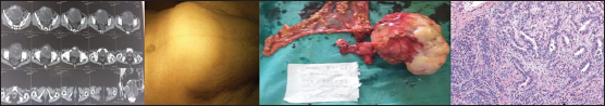
A 42-year-old woman presented Amenorrhea for 14 months. Change in her voice for 1 year and Excessive hair growth on her face, chest, and limbs for the last 2 months. She complained of vague abdominal discomfort. No history of anorexia, weight loss, increased libido. Her medical and family history was unremarkable. On examination - Hirsutism and clitoromegaly. Lump of size 10×8 cm palpable in left iliac fossa. Vaginal examination revealed a firm and mobile cystic mass in the right adnexa. An ultrasound examination of the pelvis showed a 17 × 13 × 9-cm heterogeneous solid cystic mass replacing the left ovary. The right ovary and the uterus were normal. CECT Scan Abdomen-Large heterogenous encapsulated solid soft tissue mass lesions containing areas of calcification arising from left ovary of size 17 × 13 × 10.6cm causing displacement of urinary bladder and surrounding bowel loops. Serum testosterone level -2 ng/mL (normal, 0.2–1.2 ng/mL); (DHEAS), CA 125, and alpha fetoprotein (AFP) -normal. On Laparotmy-Large mass of size 17 × 13 cm arising from left adnexa. Uterus and right ovary grossly normal. Total Abdominal hysterectomy, B/L Salpingo-opherectomy and infracolic omentectomy was done. Peritoneal washing were sent for cytologic examination for malignant cells. No liver metastasis. The post operative period was uneventful. Histopathology revealed- confirmed it be Sertoli Leydig cell tumor. 3month follow up – resolution of her virilization symptoms. No increase of her hirsutism. Repeat testosterone levels - within normal range.
Conclusion:
Only few cases of SLCT have been reported till date Prognosis depends on extent of disease, stage of disease, tumour differentiation, grade. The treatment should be individualized according to the location, state of spread and the patient's condition.