

Translate this page into:
Step-by-Step Guide to Surface Mold Applicator-Based Brachytherapy in a Case of Early Carcinoma Penis: An Organ-Preserving Approach
Address for correspondence Asha Ranjan, MD, Department of Endocrinology, Institute of Medical Sciences, Banaras Hindu University, Room No. 33, Nagarjuna Hostel, BHU, Varanasi, Uttar Pradesh, 221005, India. asharjn.85@gmail.com
This article was originally published by Thieme Medical and Scientific Publishers Pvt. Ltd. and was migrated to Scientific Scholar after the change of Publisher.
Abstract
Purpose In this report we discuss an individually customized surface mold applicator technique to treat a case of early carcinoma penis. This case report is one of its kind which describes the brachytherapy treatment planning based on orthogonal X-ray technique.
Material and methods T1N0 disease with squamous cell carcinoma histology involving the foreskin of penis and abutting the glans penis was treated by surface mold applicator technique. Impression of the organ was taken using alginate. Cast was prepared from the impression using Type 4 gypsum. The acrylic mold-based applicator was fabricated on the cast. The catheters were positioned and fixed on the acrylic mold. The organ is placed in the mold-based applicator and the catheters are reconstructed on two-dimensional imaging. The brachytherapy is delivered by the 18-channel high-dose rate Oncentra brachytherapy using Ir192 source. The dose given was 45 Gy in 15 fractions, twice daily more than 6 hours apart.
Results Patient was reviewed after 6 weeks for follow-up and there was complete regression of the ulcer. At present, patient is on follow-up for the last 10 months and is now disease-free.
Conclusion With judicious patient selection, surface mold brachytherapy is an attractive alternative to the interstitial brachytherapy. It is comfortable for the patient, is easily repeatable, and is time saving in high work load and limited resource settings. Early results with this technique are promising.
Keywords
brachytherapy
carcinoma penis
surface mold applicator

Introduction
Surgical options currently available for the primary tumor involving penis are local tumor excision, partial, or total penectomy.1 Though partial or total penectomy guarantees excellent local control rates, such radical surgical approaches are invariably associated with impairment of sexual function and quality of life and can have a devastating effect on a man's self-image.2 There are reports of suicide also after this procedure. Novac et al found increased anxiety and depression in the group with total penectomy.3 The vast majority (95%) of penile malignancies are squamous cell carcinomas (SCCs) and these are known to be radiosensitive.4 External beam radiotherapy (EBRT) and brachytherapy (BT) are both used in the primary treatment of T1–2 penile cancers < 4 cm in size either separately or together.5,6 The different types of BT used are external isotope mold, low dose rate BT, pulse dose rate BT, or high dose BT.7 The type of radiotherapy (RT) best suited for a patient depends upon the tumor location, size, thickness, and its proximity to the urethra. In well-selected patients with T1–2 tumors (lesser than 4 cm), organ preservation can be achieved with primary tumor RT. The actuarial penile preservation rate after BT was 87% at 5 years.8 EBRT has universal applicability and can be used in all RT departments, whereas BT needs expertise. Organ-sparing local tumor excision when combined with RT can achieve good local control.9 Surgery can always be an option as a salvage procedure.10,11 In a superficial tumor, the fibrosis, urethral stenosis, and sexual dysfunction usually associated with interstitial BT can be avoided by using the surface mold applicator technique.7,12,13 These surface mold applicators need to be individually customized depending on the location and extent of disease.
In this report, we discuss an individually customized surface mold applicator technique to treat a case of early carcinoma penis. This case report is unique in a way that it describes planning based on orthogonal X-ray and which can be a useful guide in a limited resource and high patient burden setting.
Case Report
A 65 years old male underwent circumcision for the ulcer over the foreskin which he had noticed 5 months back. Two months later, a recurrent ulcer developed at the circumcision site. There was no history of pain, active bleeding, or pus discharge from the ulcer. On examination, there was an ulcerative growth at the prepucial skin on ventral aspect of size 2 × 2 cm, reaching up to glans penis with no active bleeding or pus discharge from the ulcer. There was no palpable lymphadenopathy present in the inguinal region. Histopathology of the circumcision specimen revealed moderately differentiated SCC involving subepithelium with no lymphovascular and perineural invasion. The invasive tumor was 0.2 cm away from the closest mucosal margin. Deep margin was free of tumor. Positron emission tomography scan revealed mild focal fluorodeoxyglucose uptake (standardized uptake value max 3.4) at the postoperative site. There was no lymphadenopathy. In view of T1a lesion, treatment options were discussed with the patient. He declined for resurgery and opted for BT. In T1G2 tumors, risk of occult lymph node metastases is 30 to 35%, but in view of absence of vascular or lymphatic invasion and superficial growth pattern, surveillance is recommended for lymph node management.5 Therefore, only the primary lesion was treated by surface mold applicator BT. Mold-based applicator was prepared in consultation with the Department of Prosthodontics, HP Government Dental College and Hospital, Shimla, Himachal Pradesh, India. The following procedure was followed:
-
Impression of the organ was taken using alginate (hydrocolloid impression material).
-
Cast was prepared from the impression using Type 4 gypsum.
-
On the cast thus obtained, the acrylic mold-based applicator was fabricated.
-
The position of catheters was planned with each catheter placed parallel and 1 cm apart.
-
The catheters were positioned and fixed on the acrylic mold.
-
The organ is placed in the mold-based applicator, fixed with the help of temporary adhesive tapes, and orthogonal X-ray is taken at the conventional simulator.
-
The X-ray images were transferred from the conventional simulator to the Oncentra BT treatment planning system.
-
The catheters were reconstructed on two-dimensional imaging and treatment prescribed at 0.5 cm from the inner surface of the mold. Scrotal shield was kept in position, while delivering the treatment.
-
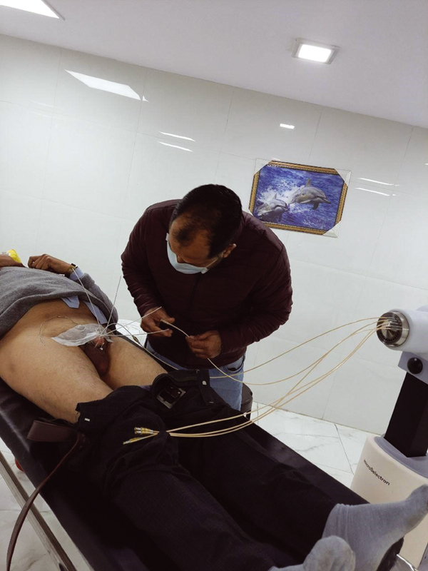
The BT was delivered by the 18-channel high-dose rate (HDR) Oncentra BT using Ir192 source (Fig. 1).
-
Fig. 1 Treatment delivered by the 18-channel high-dose rate (HDR) Oncentra brachytherapy using Ir192 source.
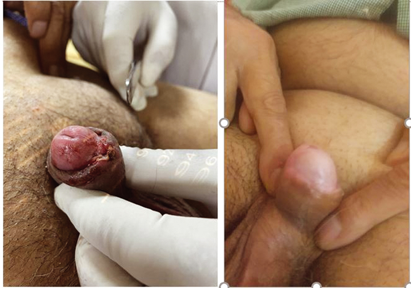
While planning, care was taken to avoid overlapping of 200% isodose lines to prevent skin necrosis and also to avoid cold spots in the treatment area. The dose given was 45 Gy in 15 fractions, twice daily more than 6 hours apart. Patient was reviewed after 6 weeks for follow-up and there was complete regression of the ulcer (Fig. 2). At local site, only mild scarring and hypopigmentation was noted at the treated area and patient is on follow-up for the last 10 months and is disease-free.
-
Fig. 2 Penile ulcer at diagnosis and at first follow-up (complete response of primary disease).
Discussion
Two types of BT techniques are widely used to treat early carcinoma penis: interstitial–invasive, using catheters and needles and surface mold BT and noninvasive and individualized applicator-based approach. Patients should be judiciously selected for the second approach. T1N0, grade 1 and 2 tumors are best suited for surface mold BT. The size of tumor and depth of invasion are the factors predicting local control.5 The ideal tumor for BT should be less than 4 cm in maximum diameter with less than 1 cm depth of invasion.5,6 Of these, in our institution, we prefer surface mold BT for tumors with depth of invasion less than 0.5 cm. Case series by Dee et al demonstrates that it can also be applied to myriad metastases, as a palliative procedure, expanding the role for penile BT.12 As urethra receives minimal dose contribution from the sources, the urethral stricture is not a concern.
Interstitial BT can be offered to a greater subset of patients which include T1, T2, and selected T3 SCC patients.13 It requires hospital admission and is always done under general anesthesia or spinal anesthesia. Also, being an invasive procedure, it will require vigorous pain management during the treatment duration. Urethral strictures are a known late complication of interstitial BT. Urethral stenosis is reported in 10 to 45% of patients and tends to occur later in the follow-up period but usually before 3 years.7,13 There are few reports showing good results of BT in well-selected early cases of carcinoma penis.12,13,14 Dee et al presented a case series of 12 patients treated by surface mold technique.12 They used customized wrapper applicators, which were fit circumferentially and longitudinally around the penile shaft using super flap bolus material. The results were rewarding with eight patients considered disease-free. Saldi et al reported a series of 7 patients treated by customized applicators constructed using a three-dimensional printer or thermoplastic mask.14 Dose delivered was 57 Gy in 19 fractions. All patients achieved complete remission and none developed grade 3 or grade 4 acute or late toxicities. Thus, surface HDR BT in penile cancers is associated with high tolerance profile and good outcome. Crook reported results for 49 men with SCC of penis treated with primary penile interstitial BT.13 At 5 years, actuarial overall survival was 78.3% and cause-specific survival 90%. Five-year penile preservation is achieved in 86% of men without sacrifice of cancer control. The choice of treatment offered to penile cancer patients vary widely in different institutes. No randomized control trials presently exist to compare the results of different treatment modalities available. There is extensive data regarding cure of the disease after surgery in penile cancers. But, quality of life, psychological status, and sexual health issues after such surgery is rarely studied. With more patients treated by penile preservation approach, this aspect of the patient's health should also be studied and compared with those treated by surgery. Local failures are readily salvaged by surgery but careful long follow-up is required as local failures can occur years after treatment. Mazeron et al reported that of the 11 local failures in their series of 50 patients treated by interstitial BT, only 36% were during the first 2 years, whereas 45% occurred between years 2 and 5 and 18% between years 5 and 8.6 The surface mold technique is used in our patient, as the disease was early and localized. Thus, this treatment was easily performed on an outpatient basis, did not require anesthesia, pain management, was comfortable for the patient, and was easily repeatable. Planning was done on orthogonal X-rays in a conventional simulator. This procedure is time saving in high work load and limited resource settings. This modality offers good local control with a high probability of sexual function preservation and no treatment-related psychological effects. The advantages offered by surface mold BT, improves the patient compliance. Salvage surgery remains a readily available option in case of recurrence, and can regain the local control, therefore close and regular follow-up is required.15
Conclusion
With judicious patient selection, surface mold BT is a less invasive and an attractive alternative to interstitial BT.
Acknowledgment
None.
Conflict of Interest
None declared.
References
- Role of radiation therapy in the treatment of carcinoma of the penis. Br J Urol. 1994;74(05):646-651.
- [Google Scholar]
- Sexual function after partial penectomy for penile cancer. Urology. 2005;66(06):1292-1295.
- [Google Scholar]
- Psychological/psychiatric trauma in patients with penile cancer and partial or total penectomy. Arch Biol Sci. 2013;65(04):1293-1298.
- [Google Scholar]
- Cancer control and quality of life following anatomical radical retropubic prostatectomy: results at 10 years. J Urol. 1994;152:1831-1836. (5 Pt 2):
- [Google Scholar]
- Iridium-192 implantation for node-negative carcinoma of the penis: the Cookridge Hospital experience. Clin Oncol (R Coll Radiol). 2000;12(01):25-31.
- [Google Scholar]
- Interstitial radiation therapy for carcinoma of the penis using iridium 192 wires: the Henri Mondor experience (1970-1979) Int J Radiat Oncol Biol Phys. 1984;10(10):1891-1895.
- [Google Scholar]
- Interstitial brachytherapy for penile carcinoma: a multicentric survey (259 patients) Radiother Oncol. 1995;36(02):83-93.
- [Google Scholar]
- Radiation therapy alone or combined surgery and radiation therapy in squamous-cell carcinoma of the penis? Eur J Cancer. 2001;37(02):198-203.
- [Google Scholar]
- The role of brachytherapy in organ preservation for penile cancer: a meta-analysis and review of the literature. Brachytherapy. 2015;14(04):517-524.
- [Google Scholar]
- Palliative therapy for recalcitrant cutaneous T-cell lymphoma of the hands and feet with low-dose, high dose-rate brachytherapy. JAMA Dermatol. 2015;151(12):1354-1357.
- [Google Scholar]
- Surface applicator high-dose-rate fractionated brachytherapy for superficial cancers of the penis: a single-center case series and national database comparison. J Am Acad Dermatol. 2021;84(01):168-172.
- [Google Scholar]
- Contemporary role of radiotherapy in the management of primary penile tumors and metastatic disease. Urol Clin North Am. 2016;43(04):435-448.
- [Google Scholar]
- High-dose-rate brachytherapy with surface applicator in penile cancer. Brachytherapy. 2021;20(04):835-841.
- [Google Scholar]
- Penile cancer brachytherapy HDR mould technique used at the Holycross Cancer Center. J Contemp Brachytherapy. 2011;3(04):224-229.
- [Google Scholar]


