

Translate this page into:
Primary Squamous Cell Carcinoma of Stomach: A Rare Presentation
Address for correspondence Manish Sharma, MBBS, DNBR. No. 2258, Department of Medical Oncology OPD, Second Floor, Rajiv Gandhi Cancer Institute and Research Center, Sector 5, Rohini, New Delhi 110085, India. itsdrmanish@gmail.com
This article was originally published by Thieme Medical and Scientific Publishers Private Ltd. and was migrated to Scientific Scholar after the change of Publisher.
Abstract
Primary gastric squamous cell carcinoma is a very rare presentation. Its pathogenesis is obscure, and the treatment strategy is largely unknown and controversial. We report a case of primary squamous cell carcinoma of the stomach with liver metastases in a 66-year-old man. The patient presented with a 2-month history of abdominal pain, vomiting, hematemesis, and weight loss. Endoscopic examination revealed large ulceroproliferative growth in body of stomach. Integrated PET CT (positron emission tomography computed tomography) scan revealed metabolically active nodular wall thickening involving body of stomach with liver metastases. Multiple biopsies of the lesions revealed squamous cell carcinoma of the stomach. The patient was started on palliative chemotherapy and is presently in partial response to treatment. He is tolerating treatment with no major side effects.
Keywords
stomach cancer
gastric cancer
squamous cell carcinoma

Introduction
Primary gastric squamous cell carcinoma is a very rare presentation. Its pathogenesis is obscure, and the treatment strategy is largely unknown and controversial. Here we report a case of primary squamous cell carcinoma of the stomach with liver metastases in a 66-year-old man.
Case Report
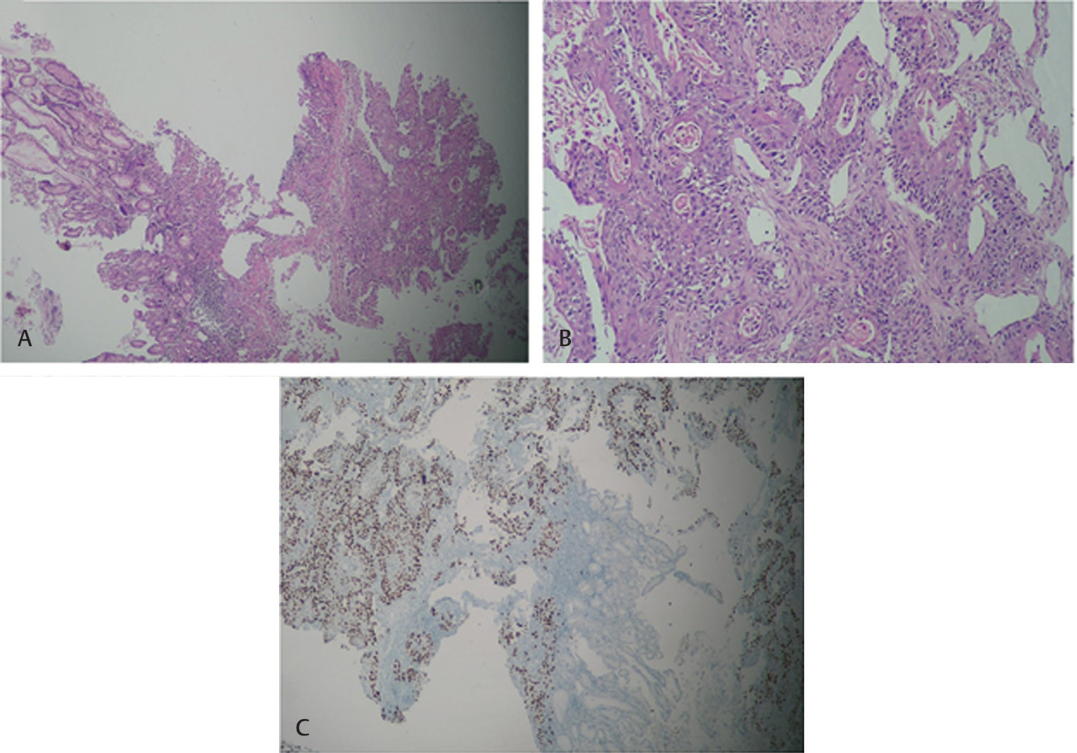
A 66-year-old man, known hypertensive and diabetic on treatment for past 20 years, presented to our hospital in January 2019 with history of abdominal pain, recurrent anemia requiring blood transfusions, vomiting, hematemesis, and weight loss. He was a chronic smoker with history of smoking 1 pack of cigarettes per day for 30 years and occasional alcohol intake. Physical examination revealed mild liver enlargement and a left supraclavicular lymph node enlargement. Laboratory tests revealed normocytic normochromic anemia (Hb = 8.3 g/dL), mild elevation of liver enzymes GGT = 105 UI/L (normal values <55 UI/L), ALP = 297 UI/L(normal values <250 UI/L), ALT = 100 UI/L (normal values <40UI/L). Tumor markers levels were CEA = 86 IU/mL, and CA19–9 = 12 IU/mL. Upper GI endoscopy revealed a large ulceroproliferative growth involving the body of the stomach. The esophagus was not involved as seen by a normal gastroesophageal junction and Z-line (Fig. 1). Multiple biopsies of the mass were taken and histological examination with immunohistochemistry revealed a moderately differentiated squamous cell carcinoma (SCC), p40+ with keratinization (Fig. 2). There was no evidence of glandular differentiation. Integrated PET CT (positron emission tomography computed tomography) scan revealed metabolically active nodular wall thickening involving body of stomach along greater and lesser curvature with metabolically active perigastric lymph nodes and metabolically active hypodense lesion in liver. Bronchoscopy and direct laryngoscopy did not reveal any evidence of the disease. Therefore, a probable metastatic primary SCC of the stomach was thought off. Since patient already had metastatic disease we did not perform gastrectomy and don’t have complete specimen of the stomach. The patient was started on paclitaxel carboplatin–based palliative chemotherapy. Post three cycles of treatment response assessment by repeat UGI endoscopy (Fig. 1) and PET CT scan, and revealed partial remission and post six cycles of treatment he had further response, but he developed grade 2 sensory neuropathy. At the time of writing this report patient continues to have stable disease 2 months after completion of treatment.

-
Fig. 1 (A) Large ulceroproliferative growth involving body stomach. (B) Normal gastroesophageal junction and Z-line.
-
Fig. 2 (A) Gastric mucosa infiltrated by squamous cell carcinoma. (B) High power view of slide 1. (C) Tumor cell express p40 on immunohistochemistry.
Discussion
Primary SCC of the stomach is extremely rare disease, with an incidence ranging between 0.04 and 0.09%. Less than 100 cases have been reported in literature.1 Diagnostic criteria were proposed by the Japanese Classification of Gastric Carcinoma; these are as follows: SCC cells, absence of adenocarcinoma components in any of the sections, and SCC arising from the gastric mucosa.2 Immunohistochemistry examination should be done on tissue samples and include CK5/6 and p40 or p63 immunomarkers.3 P40 was positive in our case with immunohistochemistry. The pathogenesis of this entity remains elusive. Various pathological mechanisms have been proposed for this entity: heterotopic squamous epithelium, squamous metaplasia, multipotent stem cells which differentiate into cells of any type, overgrowth of the squamous component in a primary adenocarcinoma, and local extension or metastasis of esophageal SCC.4 Only 56 cases of primary SCC of the stomach have been reported in Japan with a median age of 64.7± 1.7 years (29–81 years) and a male predominance. The upper third of the stomach was most common tumor location (57.1%), followed by the lower third (21.4%) and the middle third (19.6%). Tumor diameter was 2.1 to 13 cm (mean, 6.6 ± 0.3 cm).4 There is no particular management strategy and prognosis is difficult to predict. Surgical resection (R0 = no residual tumor) remains the mainstay of the treatment. In one of the case reports survival period of >3 years, with a good quality of life was reported after adjuvant chemoradiotherapy.5 Our patient is presently in partial response to palliative chemotherapy and continues to have good quality of life.
Conclusion
SCC is very rare. Its pathogenesis, diagnosis, and treatment have been considered poor. This case was presented with gastric mass with liver metastasis. Diagnosis was confirmed by histological examination and immunohistochemistry. Patient was treated with palliative chemotherapy and presently continues to be in partial response to treatment.
Conflict of Interest
None declared.
References
- Primary adenosquamous carcinoma of the stomach. A case report and review. Cancer. 1969;24(05):985-995.
- [Google Scholar]
- Japanese classification of gastric carcinoma: 3rd English edition. Gastric Cancer. 2011;14(02):101-112.
- [Google Scholar]
- Primary squamous cell carcinoma of the stomach. Report of a case and review of literature. Hepatogastroenterology. 2001;48(40):1033-1036.
- [Google Scholar]
- Primary squamous cell carcinoma of the stomach: A case report. Oncol Lett. 2014;8(05):2122-2124.
- [Google Scholar]
- Squamous cell carcinoma of the celiac area. Report of a case and review of the literature [in French] Gastroenterol Clin Biol. 2002;26(12):1168-1171.
- [Google Scholar]


