Translate this page into:
Ocular surface squamous neoplasia in Ibadan, Nigeria
This article was originally published by Thieme Medical and Scientific Publishers Private Ltd. and was migrated to Scientific Scholar after the change of Publisher.
Abstract
Introduction: Squamous cell carcinoma is the most common malignancy of the conjunctiva worldwide. Ocular surface squamous neoplasia (OSSN) describes the spectrum of ocular surface intraepithelial neoplasia, pre-invasive and invasive squamous cell carcinoma.
Method: This nonrandomized study aims to describe the epidemiology, clinical features and evaluate the outcome of treatment in patients with histological diagnosis of OSSN managed at a single tertiary center in Ibadan, Nigeria.
Result: Twenty-five patients were managed within the study period with a mean age of 42 ± 15.3 years and male: female ratio of 1:1.5. All patients presented with growth and redness, and, visual impairment was observed in seven (28%) patients. Fifteen (60%) patients were seropositive for HIV infection and one patient (4%) had xeroderma pigmentosum. The right side was involved in 11 (44%) patients and there were no bilateral lesions. Morphologically, 18 (72%) lesions were gelatinous, six (24%) were leucoplakic while one (4%) was nodular. Twenty-two (88%) patients underwent surgical excision with alcohol kerato-epitheliectomy and cryotherapy, while three (12%) patients had lid sparing orbital exenteration. The three (12%) patients with intraepithelial neoplasm, and six (24%) who had SCC but with tumor-free margins received no adjuvant treatment post-operatively, while 13 (52%) with SCC and microscopic margin involvement were treated with four courses of 0.04% topical mitomycin C (MMC) and the three (12%) patients who had orbital exenteration were referred for radiotherapy. The average follow-up period was 12 months, no patient was lost to follow-up and none has had recurrence.
Conclusion: OSSN occurs in younger individuals, and is strongly associated with HIV infection in our environment. Early diagnosis and intervention can prevent severe ocular morbidity. Wide surgical excision with intra-operative cryotherapy and adjuvant treatment with topical MMC post-operatively seem to give good outcome in our patients.
Keywords
Ibadan
mitomycin C
Nigeria
ocular surface squamous neoplasia
squamous cell carcinoma
Introduction
Although conjunctival invasive squamous cell carcinoma (SCC) is the most common malignancy of the conjunctiva in the world and in Sub-Saharan Africa,1,2,3 it remains an uncommon disease worldwide with wide range in the incidence of 0.02–3.5/100,000/year.1 It accounts for 4–29% of orbito-ocular tumors,4 and the third most common ocular tumor in adults.5 Recently, the terminology ocular surface squamous neoplasia (OSSN) was used to describe the spectrum of ocular surface intraepithelial neoplasia, preinvasive, and invasive SCC.5
OSSN had been noted to occur at younger age in patients with human immunodeficiency virus (HIV) infection, probably due to reduced immune response and surveillance,6 and xeroderma pigmentosum.7 Ultraviolet-light-induced DNA damage with delayed or failure of repair is thought to lead to somatic mutations and development of cancerous cells in patients with xeroderma pigmentosum.5 Other factors proposed in the etiology of OSSN include ultraviolet-B radiation,8,9,10 human papillomavirus (types 16 and 18) infection,11,12 ocular surface injury and chemical exposure,13 Vitamin A deficiency,14 exposure to petroleum products and heavy cigarette smoking,15 Caucasian ancestry,16 older age,5 and male gender.16,17
OSSN commonly start at the interpalpebral area of perilimbal conjunctiva and had been described macroscopically as gelatinous, leukoplakic, nodular or diffusely invasive.18 They are regarded as slowly growing tumors, and factors associated with recurrence include severe grades and status of the margins at excision.19 However, orbital or intraocular spread occurs in delayed presentation,20,21 and orbital involvement at the presentation is common in Sub-Saharan Africa.2,20
Treatment modalities for OSSN include wide surgical excision, adjunctive cryotherapy, topical or systemic chemotherapy with various agents and radiotherapy.22,23 Simple surgical excision without adjunctive treatment had been associated with higher rates of recurrence.19
Studies on OSSN are few in Nigeria and are retrospective in design using varying management modalities. A previous retrospectively designed study20 from this center detailed the histopathological features of these tumors, but the clinical presentation was not adequately addressed. To fully describe the epidemiology of these tumors and evaluate the clinical features and treatment outcome under a uniform treatment protocol, all consecutively managed cases within the study period were analyzed.
Methods
A total of 25 patients with histological diagnosis of OSSN, either intraepithelial or invasive SCC, managed between January 2009, and December 2012, at the Ocular Oncology Unit of the Eye Department, of a Tertiary Center in Ibadan, Nigeria, were included in the study. Data collected and analyzed from the patients included their demographics, presenting symptoms and duration, possible etiological factors, clinical features, tumor site, histological diagnosis and tumor margin control, treatment modalities and outcome, and follow-up period.
All patients with suspicious lesions had wide surgical excision using the “no touch technique”24 with the removal of 4 mm clinically tumor-free conjunctival skirt peripheral to the tumor and a thin sclera flap underneath the lesion. The corneal epithelium 2 mm anterior to the tumor was treated with absolute alcohol, scraped toward the lesion, and sharply dissected off the limbus, following which a double freeze-thaw cryotherapy was applied to the cut conjunctival edge and base of the tumor. Primary closure of the wound was done with 8/0 vicryl suture. Patients with orbital infiltration had lid sparing orbital exenteration done. All surgeries were performed by the author. Specimens were sent for histological diagnosis and tumor margin control. Patients with microscopic margin involvement received four courses of 0.04% topical mitomycin C (MMC). Each cycle consisted of 4 weeks of active treatment followed by 4 weeks of rest. Patients instilled topical MMC 4 times daily on 4 consecutive days followed by a 3-day break during each of the 4 weeks of treatment. Ethical approval was obtained, and the methods in the study adhered to the tenets of the Declaration of Helsinki.
Results
The patients’ demographics are shown in Figure 1. There were 25 patients (40% male) with a mean age of 42 ± 15.3 years. All patients were Nigerians, 22 (88%) of who had invasive SCC and three had intraepithelial neoplasia. The right side was involved in 11 (44%) patients, and there were no bilateral lesions. The lesion was sited at the nasal limbus in 11 (44%) patients, temporal limbus in 8 (32%), superior limbus in 1 (4%), and inferior limbus in 1 (8%) patients. Three (12%) patients presented with orbital involvement Figure 2 making the site of origin on the ocular surface difficult to assess. Morphologically, 18 (72%) lesions were gelatinous Figure 3, 6 (24%) were leukoplakic Figure 4, while 1 (4%) was nodular Figure 5. The most common symptoms at presentation were “growth” (100%) and “redness” (100%) Table 1. The vision was impaired by the growth in 7 (28%) patients, two of who had visual acuity of 6/24 and the others had visual acuity of <3/60. A total of 15 (60%) patients were found to be seropositive for HIV infection, and one patient (4%), the youngest case, presented with features of xeroderma pigmentosum Figure 4. A total of 10 (40%) patients were referred from other hospitals with recurrent growths having undergone simple tumor excision. Eight of them were seropositive for HIV infection.
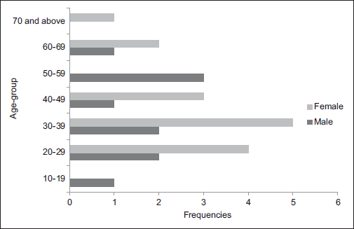
- Age and sex distribution of patients with ocular surface squamous neoplasia
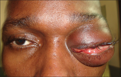
- Clinical picture of a patient with orbital infiltration
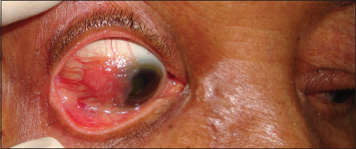
- Clinical picture of a patient with gelatinous lesion
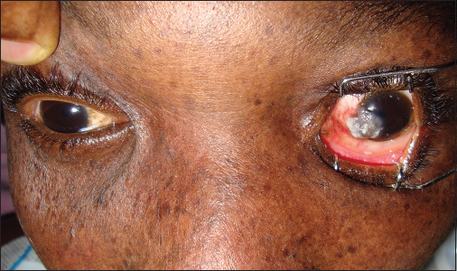
- Clinical picture of the patient with xeroderma pigmentosum showing leukoplakic lesion
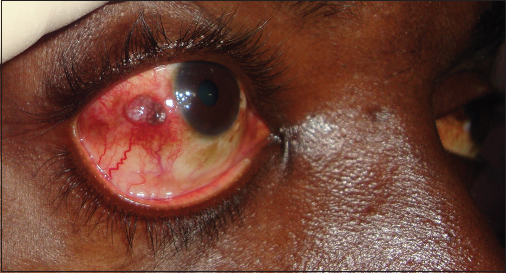
- Clinical picture of the patient with nodular lesion
|
Symptom |
Frequency |
Percentage |
|---|---|---|
|
Growth |
25 |
100 |
|
Redness |
25 |
100 |
|
Poor/reduced vision |
7 |
28 |
|
Pain |
6 |
24 |
|
Foreign body sensation |
4 |
16 |
|
Contact bleeding |
2 |
8 |
|
Pigmented skin lesions |
1 |
4 |
|
Oral sores |
1 |
4 |
*More than one option possible. OSSN - Ocular surface squamous neoplasia
There were 22 (88%) patients underwent wide surgical excision with alcohol kerato-epitheliectomy and cryotherapy, whereas 3 (12%) patients had lid sparing orbital exenteration. Three (12%) patients had intraepithelial neoplasm (moderate dysplasia), and 22 (88%) had invasive SCC. The 3 (12%) patients with intraepithelial neoplasm and 6 (24%) who had SCC with microscopic tumor-free margins received no adjuvant, topical treatment postoperatively, whereas 13 (52%) with microscopic margin involvement were treated with four courses 0.04% topical MMC. No complications were seen in patients treated with topical MMC. The 3 (12%) patients who had orbital exenteration were referred for radiotherapy. No recurrence had been recorded.
The follow-up period ranged between 5 and 46 months with an average of 12 months. No patient was lost to follow-up, and three of the patients had been followed up for more than 24 months.
Discussion
This series has shown that OSSN is not an uncommon disease in Ibadan with 25 cases managed at a single institution during a 4-year period when compared with the previous report20 of 46 cases over 15 years from three institutions in the same geographical location. This could be attributed to the prospective nature of the study with inclusion of all cases presenting during the study period, or it might be a referral bias, being a major ocular oncology center. However, there could have been a true increase in the number of cases of OSSN in the population.
The small number of intraepithelial lesions in this series, similar to a previous report,20 but at variance with reports from other countries10,17 may be due to referral bias, as 10 of the patients were referred with recurrent lesions after simple excisions from other centers. The mean age of the patients in this study is lower than previously reported in the country20 and other countries.18,25,26 The younger age of affectation in a previous study20 was attributed to a possibly shorter life expectancy in the country resulting in fewer individuals living old enough to develop the disease. However, this study has demonstrated a strong association between OSSN and patients with HIV infection who are in their younger ages. Furthermore, awareness about the disease had increased over the years, and conjunctival SCC is now considered a marker for HIV/AIDS disease in Sub-Saharan Africa.6,27,28 About two-third of the patients in this study tested positive for HIV higher than reported by Ogun et al.20 However, their inability to find an association between OSSN and HIV infection could be due to the retrospective design of their study, with a significant number of the studied cases not screened for the virus. HIV infection and xeroderma pigmentosum had been associated with higher prevalence and earlier presentation of OSSN,6,7,29 and the youngest case in this series had dermatological features of xeroderma pigmentosum.
OSSN is regarded as a low-grade malignancy10,30 and has a wide duration of symptoms before the presentation.5,25 The mean duration of symptoms in this study of 16.7 ± 7 months was higher than some studies5,25 yet with few cases of orbital tumor infiltration. In addition, there were no cases of intraocular tumor spread at variance with previous studies;6,18,20 thus, supporting the low-grade malignancy of this group of tumors.
The higher prevalence of OSSN in females in Nigeria, at variance with reports from industrialized countries,10,17,25,26 had been attributed to the sociocultural practices in the country.20 However, this might also be due to the high number of HIV-seropositive patients in this study being females.
We could not substantiate any association between OSSN and other etiologic risk factors such as smoking and exposure to petroleum products as previously suggested in literature.15 Furthermore, in line with the findings of Ogun et al.,20 no association was found with cutaneous and visceral malignancies at variance with reports from some industrialized countries.5,10,18
A large proportion of our cases was referred from other centers with recurrent lesions. These patients had undergone simple tumor excision without intraoperative cryotherapy and did not receive postoperative adjuvant chemotherapy. Simple tumor excision without cryotherapy had been associated with recurrences ranging between 15% and 52%.5,10 Other factors associated with tumor recurrence include large tumor size, positive surgical margin, increased age, and elevated proliferative index measured by Ki-67.18,25 We did not record any recurrence in this series suggesting that topical MMC in conjunction with intraoperative cryotherapy is effective in the control of microscopic tumor residual after wide excision in a Nigerian population as reported by Chen et al.26 in a non-Black population. However, long-term follow-up of the patients is on to substantiate this.
Conclusion
OSSN occurs in younger individuals, and there is a strong association with HIV infection in our environment. The early diagnosis and intervention may prevent severe ocular morbidity. Wide surgical excision with intraoperative cryotherapy and adjuvant treatment with topical MMC postoperatively seem to give a good outcome in our patients. There is, however, the need for a longer follow-up of these patients.
Financial support and sponsorship
Nil.
Conflicts of interest
There are no conflicts of interest.
References
- A clinicopathological study of orbito-ocular diseases in Ibadan between 1991-1999. Afr J Med Med Sci. 2003;32:197-202.
- [Google Scholar]
- Diseases of the outer eye In: Duke-Elder S, ed. Systems of Ophthalmology. St. Louis: CV Mosby; 1985. p. :I154-9-75.:65-75.
- [Google Scholar]
- Epibulbar squamous cell carcinomas in brothers with xeroderma pigmentosa. J Pediatr Ophthalmol Strabismus. 1991;28:350-3.
- [Google Scholar]
- Risk factors in the development of ocular surface epithelial dysplasia. Ophthalmology. 1994;101:360-4.
- [Google Scholar]
- A review of the aetiology of squamous cell carcinoma of the conjunctiva. Br J Cancer. 1996;74:1511-3.
- [Google Scholar]
- Conjunctival and corneal intraepithelial and invasive neoplasia. Ophthalmology. 1986;93:176-83.
- [Google Scholar]
- Human papillomavirus DNA in tissues and ocular surface swabs of patients with conjunctival epithelial neoplasia. Invest Ophthalmol Vis Sci. 1992;33:184-9.
- [Google Scholar]
- High frequency of human papillomavirus 6/11, 16, and 18 infections in precancerous lesions and squamous cell carcinoma of the conjunctiva in subtropical Tanzania. Am J Clin Pathol. 2004;122:938-43.
- [Google Scholar]
- Bowen's disease of the conjunctiva: A misnomer In: Jakobiec F A, ed. Ocular and Adnexal Tumors. Birmingham, AL: Aesculapius; 1978. p. :553-71.
- [Google Scholar]
- Factors associated with conjunctival intraepithelial neoplasia: A case control study In: Ophthalmic Surg. 1990. p. :27-30. 21
- [Google Scholar]
- Epidemiology of squamous cell conjunctival cancer. Cancer Epidemiol Biomarkers Prev. 1997;6:73-7.
- [Google Scholar]
- Adjunctive radiotherapy with strontium-90 in the treatment of conjunctival squamous cell carcinoma. Int J Radiat Oncol Biol Phys. 1988;14:435-43.
- [Google Scholar]
- Intraepithelial and invasive squamous cell carcinoma of the conjunctiva: Analysis of 60 cases. Br J Ophthalmol. 1999;83:98-103.
- [Google Scholar]
- Late recurrences and the necessity for long-term follow-up in corneal and conjunctival intraepithelial neoplasia. Ophthalmology. 1997;104:485-92.
- [Google Scholar]
- Intraepithelial and invasive squamous neoplasms of the conjunctiva in Ibadan, Nigeria: A clinicopathological study of 46 cases. Int Ophthalmol. 2009;29:401-9.
- [Google Scholar]
- Invasive squamous cell carcinoma of the conjunctiva. Arch Ophthalmol. 1994;112:1342-5.
- [Google Scholar]
- Current treatment options for conjunctival and corneal intraepithelial neoplasia. Ocul Surf. 2003;1:66-73.
- [Google Scholar]
- Squamous cell carcinoma of the conjunctiva with orbital invasion: Orbital exenteration or minimally invasive procedure? Ophthalmologe. 2006;103:693-7.
- [Google Scholar]
- Squamous cell carcinoma of the conjunctiva: A series of 26 cases. Br J Ophthalmol. 2002;86:168-73.
- [Google Scholar]
- Mitomycin C as an adjunct in the treatment of localised ocular surface squamous neoplasia. Br J Ophthalmol. 2004;88:17-8.
- [Google Scholar]
- Carcinoma of the conjunctiva and HIV infection in Uganda and Malawi. Br J Ophthalmol. 1996;80:503-8.
- [Google Scholar]
- Xeroderma pigmentosum in three consecutive siblings of a Nigerian family: Observations on oculocutaneous manifestations in black African children. Br J Ophthalmol. 2001;85:110-1.
- [Google Scholar]


