Translate this page into:
Emergence of micronuclei and apoptosis as potential biomarker of oral carcinogenesis: An updated review
Address for correspondence: Dr. Dipak Baliram PatilPlot No 9 Dhake Wadi Jalgaon, Maharashtra, India. dipak123patil@gmail.com
This article was originally published by Thieme Medical and Scientific Publishers Private Ltd. and was migrated to Scientific Scholar after the change of Publisher.
Abstract
More than 95% cancers of oral cavity are squamous cell carcinoma. They contribute major health problems in developing countries like India. The critical etiological factor for oral squamous cell carcinoma (OSCC) is the consumption of tobacco in various forms. OSCC results from alterations in genes that control the cell cycle or that are involved in deoxyribonucleic acid repair and are characterized by the loss of ability of cells to evolve to death when genetic damage occurs. The occurrence of chromosomal damage can be evaluated by counting micronuclei (MNs) and degenerative alterations, indicative of apoptosis such as karyorrhexis, pyknosis, and condensed chromatin. Apoptosis has been associated with the elimination of potentially malignant cells, hyperplasia, and tumor progression. Hence, reduced apoptosis or its resistance plays a vital role in carcinogenesis. MNs are one of such biomarkers that are cytoplasmic chromatin masses with the appearance of small nuclei that arise from lagging chromosomes at anaphase or from acentric chromosome fragments. They are induced in the cells by numerous genotoxic agents that damage the chromosome. Bigger MNs result from exclusion of whole chromosome following damage to the spindle apparatus of the cell (aneugenic effect), whereas smaller MNs result from structural aberrations causing chromosomal fragments (clastogenic effect). Thus, MN count and apoptosis can be a useful biomarker, and it can be used as a screening test for patients with habit of tobacco consumption and patients with manifestations of oral lesion including premalignant and malignant conditions.
Keywords
Apoptosis
chromosomal damage
genotoxic damage
human micronucleus project on exfoliated buccal cells
micronucleus
reactive oxygen species production
Introduction
Oral cancer is among the ten types of malignant neoplasia of highest incidence worldwide and is particularly common in developing countries.1 More than 95% of carcinomas of the oral cavity are of squamous cell type. They constitute a major health problem in developing countries, representing a leading cause of death. The survival index continues to be small (50%) as compared to the progress in diagnosis and treatment of other malignant tumors. According to the World Health Organization, carcinoma of oral cavity in males in developing countries is the sixth most common cancer after lung, prostate, colorectal, stomach, and bladder cancer, while in females, it is the tenth most common site of cancer after breast, colorectal, lung, stomach, uterus, cervix, ovary, bladder, and liver.2
Cancer occurs through multiple steps, each characterized by the sequential stimulation of additional genetic defects, followed by clonal expansion. The genetic alterations observed in head and neck cancer are mainly due to oncogene activation and tumor suppressor gene inactivation, leading to deregulation of cell proliferation and death.2 Since tissue homeostasis is the result of a subtle balance between proliferation and cell death, too little cell death by apoptosis can promote tumor formation as well as progression. Evasion of apoptosis is one of the hallmarks of human cancers that promote tumor formation and progression as well as treatment resistance.3
The discovery of apoptosis’ escape mechanisms has a great potential for translational medicine since such defects in apoptosis molecules may serve as targets for the design of novel therapeutic strategies as well as molecular markers to predict treatment response and prognosis. The enormous progress in apoptosis research has started to be translated into the development of innovative cancer diagnostics and therapeutics. Similar to other types of malignant neoplasia, oral cancer results from alterations (point mutations and chromosomal abnormalities) in genes that control the cell cycle and/or in genes that are involved in deoxyribonucleic acid (DNA) repair.
Micronuclei (MNs) are extra nuclear-cytoplasmic bodies, and they are induced in oral exfoliated cells by a variety of substances, including genotoxic agents and carcinogenic compound in tobacco, betel nut, and alcohol.4 MN detection in oral squamous cell carcinoma (OSCC) has been shown to have a sensitivity of 94%, a specificity of 100%, and an accuracy of 95%, Thus, they are good prognostic indicators and occurrences of chromosomal damage in the oral epithelium can be evaluated using the MN test, as suggested by Dórea et al.1 Previous studies have shown false-positive results in MN frequency when using Romanowsky-type stains. Studies have shown that the sensitivity of DNA-specific dyes is almost twice as compared to nonspecific dyes. Cells containing MN on the bright field can be confirmed as being positive by examining the cells under fluorescence, and the nuclear texture which is important in classifying condensed chromatin and karyorrhectic cells may be easier to discern.4
Genotoxic Damage and Biomarkers
It is generally accepted that oral carcinogenesis is a multistep process of accumulated genetic damage, leading to cell dysregulation with disruption in cell signaling, DNA repair, and cell cycle, which are fundamental to homeostasis.5 These events can be conveniently studied in the buccal mucosa, which is an easily accessible tissue for sampling cells in a minimally invasive manner and does not cause undue stress to study subjects. This method is increasingly being used in molecular epidemiological studies to investigate the impact of nutrition, lifestyle factors, genotoxic exposure, and genotype on DNA damage and cell death. Exfoliative cytology of the buccal mucosa in cases of oral cancers has recently been successfully used to identify various mutations in important genes using a polymerase chain reaction and other techniques and also to quantify formation of DNA-carcinogen adducts.6 The present goal in many research laboratories is to develop screening strategies indicating individual cancers with certain biomarkers. Biomarkers are instruments of individual tumor prevention and help detect high-risk patients. They allow statements concerning environmental and occupational exposition and further give information on the status of susceptibility. Biomarkers are divided into three groups: the first to define the exposure to carcinogenic agents, the second to show biological effects on the target tissue, and the third to give information about the individual susceptibility.7
Chromosomal Damage and Reactive Oxygen Species Production
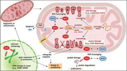
The DNA, carrier of the human genetic code, can be damaged throughout a wide range of reasons. Each of the ∼1013 cells in the human body receives thousands of DNA lesions every day. Impaired or incorrect repair due to blocked genome replication and transcription may lead to mutations or genome aberrations causing an increased risk for diverse cancers, neurodegenerative diseases, or cardiovascular diseases. DNA damage also arises by other mechanisms due to oxidative stress. Cellular processes, external factors, and/or disease states can lead to the formation of reactive oxygen species (ROS) that may interact with the DNA. On the one hand, ROS are released from phagocytes to destroy cells infected with virus or bacteria and, on the other hand, formed by ionizing and ultraviolet radiation or potent environmental carcinogens and chemicals in the mitochondria. Their negative impacts are based on an overload of O2-, HO°, or H2O2 and further lead to genomic alterations to enhanced lipid oxidation, protein oxidation, and DNA oxidation8 Figure 1.

- The impact of reactive oxygen species on deoxyribonucleic acid and proteins. Reactive oxygen species are generated in mitochondria, scavenging systems exist and if mitochondrial deoxyribonucleic acid functions well-damaged proteins get removed and replaced by new ones. In more severe cases as seen in complex IV deficiency, expression of additional genes gets stimulated to rescue lost functions. Oxidative damage furthermore leads to induction of apoptosis
Apoptosis and Cancer
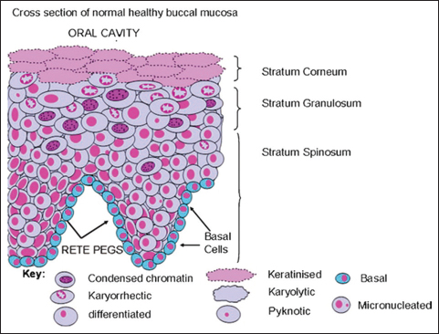
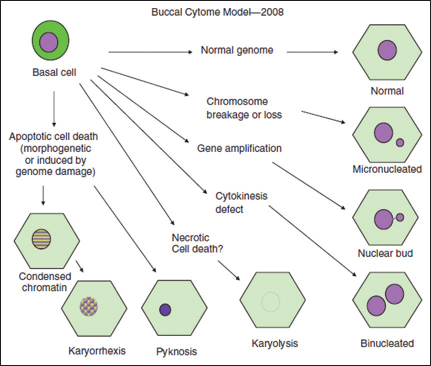
Defects in programmed cell death (apoptosis) mechanisms play an important role in tumor pathogenesis, allowing neoplastic cells to survive over intended lifespans, subverting the need for exogenous survival factors, and providing protection from oxidative stress and hypoxia as the tumor mass expands.8 Morphological hallmarks of apoptosis in the nucleus are chromatin condensation and nuclear fragmentation, which are accompanied by rounding up of the cell, reduction in cellular volume (pyknosis), and retraction of pseudopods. Chromatin condensation starts at the periphery of the nuclear membrane, forming a crescent-like or ring-like structure. The chromatin further condenses until it breaks up inside a cell with an intact membrane, a feature described as karyorrhexis (KR). The plasma membrane is intact throughout the total process. At the later stage of apoptosis, some of the morphological features include membrane blebbing, ultrastructural modification of cytoplasmic organelles, and loss of membrane integrity9 Figures 23.

- Diagrammatic representation of a cross-section of normal buccal mucosa of healthy individuals illustrating the different cell layers and possible spatial relationships of the various cell types

- Diagrammatic representation and possible inter-relationships between the various cell types observed in the buccal cytome assay based on the scheme proposed by Tolbert et al.
Apoptosis defects are now considered an important complement of proto-oncogene activation as many deregulated oncoproteins that drive cell division also trigger apoptosis (e.g., Myc, E1a, and cyclin-D1). On the other hand, the noncancerous cells have DNA repair machinery. Defects in DNA repair and/or chromosome segregation normally trigger cell suicide as a defense mechanism for eradicating genetically unstable cells, and thus, such suicide mechanism's defects permit survival of genetically unstable cells, providing opportunities for selection of progressively aggressive clones, and may promote tumorigenesis. This apoptosis’ defects may allow epithelial cells to survive in a suspended state, without attachment to extracellular matrix which facilitate metastasis.10
Micronuclei: Formation and Identification
MNs are one of such biomarkers that are cytoplasmic chromatin masses with the appearance of small nuclei that arise from lagging chromosomes at anaphase or from acentric chromosome fragments. They are induced in the cells by numerous genotoxic agents that damage the chromosome.6 The damaged chromosomes, in the form of acentric chromatids or chromosome fragments, lag behind in anaphase when centric elements move toward the spindle poles. After telophase, the undamaged chromosomes, as well as the centric fragments, give rise to regular daughter nuclei. The lagging elements are included in the daughter cells too, but a considerable proportion is transformed into one or several secondary nuclei, which are, as a rule, much smaller than the principal nucleus and are therefore called MNs.4
Bigger MNs result from exclusion of whole chromosome following damage to the spindle apparatus of the cell (aneugenic effect), whereas smaller MNs result from structural aberrations causing chromosomal fragments (clastogenic effect). MNs are induced in oral exfoliated cells by a variety of substances, including genotoxic agents and carcinogenic compound in tobacco, betel nut, and alcohol. Tobacco-specific nitrosamines have been reported to be potent clastogenic and mutagenic agents.11
The use of MNs as a measure of chromosomal damage in the peripheral blood lymphocytes was first proposed by Countryman and Heddle in 1976 and subsequently improved with the development of the cytokinesis-block MN (CBMN) method, which allowed MNs to be scored specifically in cells that had completed nuclear division.11 More direct methods include measurements in the peripheral blood lymphocytes and buccal cells. Single-gel electrophoresis (COMET assay) provides investigation of DNA single- or double-strand breaks and other oxidized DNA bases in lymphocytes and can also be adapted in buccal cells (with concern to some limitations). In both, lymphocyte and buccal cell chromosomal aberrations as MNs can be obtained and related to chromosomal damage.12
Rao et al. evaluated DNA damage levels in the peripheral blood leukocytes (PBLs) of tobacco-habituated individuals with clinically normal mucosa and patients with oral carcinoma and compared with a control group of healthy volunteers using single-cell gel electrophoresis (SCGE) and fluorescent ethidium bromide stain.13
No standardized protocol for the buccal MN cytome (BMcyt) is available to date in the peripheral blood lymphocytes in relation to CBMN cytome (CBMNcyt) assay. For solving among other topics, the issue of unification the “human MN project on exfoliated buccal cells” was launched. The project aims to refine the assay by strong collaboration with international laboratories.14
Furthermore, first approaches for automation of the BMcyt are described by Leifert et al. in 2011. Laser scanning cytometry is suggested to measure biomarkers for DNA damage (MNs, broken eggs), cytokinesis defects (binucleated cells [BNs]), or cell death (condensed chromatin, KR, pyknosis, and karyolitic cells) following standardized scoring criteria. Automation may contribute to maintaining uniform results.15
Compared to the CBMNcyt, no blood samples need to be taken for the BMcyt and buccal tissue is easily reachable, and therefore, the method serves perfectly for large biomonitoring studies, especially in pediatric population. Easy storing of samples and low costs favor this method. Since buccal tissue allows immediate assessment of DNA damage, no further replication step is required. The BMcyt is successful without establishing cell cultures for analyzing metaphase and interphase in binucleated lymphocytes. The strong correlation of MN frequency in buccal cells with MN frequencies in lymphocytes warrants great potential in biomonitoring risks for certain diseases, in the first place for cancer.14
Projects for the Apoptosis and Buccal Cell Micronuclei Assay
Rao et al.13 evaluated DNA damage levels in the PBLs with respect to clinical staging and histopathological grading of patients with oral carcinoma using SCGE and fluorescent ethidium bromide stain. They found that DNA damage levels increased from clinical Stage I to Stage IV, while considering histopathological grading, the mean DNA damage levels in leukocytes gradually increased from Grade I to Grade III. They have concluded that tobacco-using individuals are more prone to oral cancer and the effects of tobacco on PBLs, particularly lymphocytes, may possibly be derived from a further study of DNA damage in the peripheral blood cells in a group of individuals addicted to tobacco, however showing no histologic changes indicative of malignancy. Udupa et al.16 undertook a study to assess the DNA damage in oral submucous fibrosis (OSF) patient and healthy groups using COMET assay using ethidium bromide stain. The DNA damage was evaluated by measuring the tail length. They have noted an increase in tail length in the buccal epithelial cells of oral sub mucous fibrosis (OSMF) group when compared with healthy group. Furthermore, there was a significant increase in the DNA damage with duration of habits. They have suggested that measuring the extent of tail length in oral epithelium would provide direct evidence of association of DNA damage in oral epithelium with these deleterious oral habits. This assay could be incorporated into clinical practice to improve outcome and survival of persons at high risk for oral cancer.
Guttikonda et al.17 evaluated DNA damage levels in the PBLs of tobacco-habituated individuals with clinically normal mucosa and patients with oral carcinoma and compared with a control group of healthy volunteer using SCGE and fluorescent ethidium bromide stain. They found that the mean DNA damage levels in normal subjects, tobacco users with normal mucosa, and tobacco users with oral cancer patients increased significantly. They have concluded that DNA damage evaluation in PBLs by SCGE technique is a sensitive and reliable indicator of tobacco insult and OSCC.
Nersesyan et al.18 studied the effect of different staining procedures on the outcome of MN frequency investigations, in oral mucosa cells of heavy smokers and nonsmokers. MN frequencies were evaluated with nonspecific (Giemsa, May-Grunwald-Giemsa) and DNA-specific (4, 6-diamidino-2-phenylindole, i.e., DAPI, Feulgen, acridine orange) stains. They have shown that in nonspecific Giemsa-based stains, the frequencies of MNs were significantly (4–5-fold) higher in the smoker group than nonsmokers while no significant increase was observed with any of the DNA-specific stains. Furthermore, their analysis showed that MN frequencies scored in Giemsa-stained slides correlated significantly with KR, karyolysis (KL), condensed chromatin, and binucleates, whereas no such correlations were found with DNA-specific stains. These findings indicate that nuclear anomalies (and possibly keratin bodies) may be misinterpreted as MNs with nonspecific DNA stains and lead to false-positive results in studies with cells of epithelial origin. They concluded that exposure of oral mucosa cells to genotoxic carcinogens contained in tobacco smoke does not lead to induction of MNs in these cells. The increased risks of oral cancer in smokers may be due to acute toxic effects and inflammation that is not associated with MNs formation. Therefore, the use of MNs in exfoliated epithelial cells as biomarkers of exposure to lifestyle- and occupation-related genotoxic carcinogens warrants further evaluation in general.
Jyoti et al.19 evaluated frequency of MN in exfoliated buccal epithelial cells of OSF patients by acridine orange fluorescent staining and observed a significant increase in frequency of MN in OSMF patients as compared to gutkha chewers and controls. They have used acridine orange due to its fluorescent nature and easier visibility of MN present in buccal epithelial cells. They have concluded that chewing gutkha along with smoking is more dangerous for human health as it hastens the incidence of OSMF. Toy et al.20 evaluated the cytotoxicity and genotoxicity of fixed orthodontic treatment with three different light-cured orthodontic bonding composites by analyzing MN formation and nuclear alterations, such as KR, KL, and BNs, in the buccal mucosa during a 6-month period. Parameters were scored under light microscope using acridine orange stain. They have shown that MN rates did not significantly differ among different time points within the same cell type. In contrast, the number of BNs in buccal epithelial cells significantly increased in all composite groups while KL frequency significantly increased between the beginning and end of the study in the Kurasfer and Gren Gloo groups. Thus, they concluded that after 6 months of fixed orthodontic treatment with different light-cured composites, morphological signs of cytotoxicity were observed, but genotoxic effects were absent, and further investigation will be necessary to confirm and expand their findings. Bansal et al.21 detected MNs in exfoliated oral mucosal cells in individuals using various tobacco forms from the last 5 years. They have selected 25 smokeless tobacco users, 25 smokers, and 25 nontobacco users and used Papanicolaou staining technique. They found that MNs were significantly higher in smokeless tobacco users than in smokers and controls and concluded that increase in the number of MNs provides evidence that smokeless tobacco chewers and smokers may be at a high risk for developing oral cancer. In comparison, the cellular changes associated with smokeless tobacco use were more than that in smokers, thus indicating more carcinogenic potential of smokeless tobacco and MN assay can be used as a biomarker of genotoxicity and epithelial carcinogenic progression. Kamath et al.22 assessed the presence or increase of MNs in buccal smears of individuals with tobacco habits against a control group of teetotalers with using standard Papanicolaou stain. They showed that there was a direct correlation with increased MN in smokers and increased correlation of presence of MN with age, duration, and frequency of smoking. Interestingly, peak MN detection was seen in patients in the third decade and above, when effects of smoking would be considered the most. A direct correlation was also observed between the expression of MN and increased frequency of smoking (>10 cigarettes/day). They have concluded that assessment of predysplastic changes using noninvasive, easy procedure like smears can be advocated in all routine field screening, MN would be better indicators for such changes, and patients with high risk can be counseled by delivering the ill effects of tobacco accordingly.
Conclusion
Cancer can be viewed as the result of a succession of genetic changes during which a normal cell is transformed into a malignant one, while evasion of cell death is one of the essential changes in a cell that cause this malignant transformation. Apoptosis has been associated with elimination of potentially malignant cells, hyperplasia, and tumor progression. Hence, reduced apoptosis or its resistance plays a vital role in carcinogenesis. MN count and apoptosis can be a useful biomarker and it can be used as a screening test for patients with habit of tobacco consumption and patients with manifestations of oral lesion including premalignant and malignant conditions.
Financial support and sponsorship
Nil.
Conflicts of interest
There are no conflicts of interest.
References
- Chromosomal damage and apoptosis in exfoliated buccal cells from individuals with oral cancer. Int J Dent. 2012;2012:457054.
- [Google Scholar]
- Oral squamous cell carcinoma: Etiology, pathogenesis and prognostic value of genomic alterations. Indian J Cancer. 2006;43:60-6.
- [Google Scholar]
- Clinico-pathological correlation of micronuclei in oral squamous cell carcinoma by exfoliative cytology. J Oral Maxillofac Pathol. 2008;12:2-6.
- [Google Scholar]
- Prognostic and predictive factors in oral squamous cell cancer: Important tools for planning individual therapy? Oral Oncol. 2004;40:110-9.
- [Google Scholar]
- Exfoliative cytology of normal buccal mucosa to predict the relative risk of cancer in the upper aerodigestive tract using the MN-assay. Oral Oncol. 2000;36:550-5.
- [Google Scholar]
- Oxidative DNA damage: Mechanisms, mutation, and disease. FASEB J. 2003;17:1195-214.
- [Google Scholar]
- Apoptosis and molecular targeting therapy in cancer. Biomed Res Int. 2014;2014:150845.
- [Google Scholar]
- Chromosome 17 and 21 aneuploidy in buccal cells is increased with ageing and in Alzheimer's disease. Mutagenesis. 2008;23:57-65.
- [Google Scholar]
- Comparison of DNA damage and repair following radiation challenge in buccal cells and lymphocytes using single-cell gel electrophoresis. Int J Radiat Biol. 2004;80:517-28.
- [Google Scholar]
- Single cell gel electrophoresis on peripheral blood leukocytes of patients with oral squamous cell carcinoma. J Oral Pathol Med. 1997;26:377-80.
- [Google Scholar]
- The HUman microNucleus project on eXfoLiated buccal cells (HUMN (XL)): The role of life-style, host factors, occupational exposures, health status, and assay protocol. Mutat Res. 2011;728:88-97.
- [Google Scholar]
- Automation of the buccal micronucleus cytome assay using laser scanning cytometry. Methods Cell Biol. 2011;102:321-39.
- [Google Scholar]
- The comet assay a method to measure DNA damage in oral submucous fibrosis patients: A case-control study. Clin Cancer Invest J. 2014;3:299-5.
- [Google Scholar]
- DNA damage in peripheral blood leukocytes in tobacco users. J Oral Maxillofac Pathol. 2014;18:S16-20.
- [Google Scholar]
- Effect of staining procedures on the results of micronucleus assays with exfoliated oral mucosa cells. Genet Mol Res. 2006;5:45-54.
- [Google Scholar]
- Evaluation of micronucleus frequency by acridine orange fluorescent staining in bucccal epithelial cells of oral submucosus fibrosis (OSMF) patients. Egypt J Med Hum Genet. 2013;14:189-93.
- [Google Scholar]
- Evaluation of the genotoxicity and cytotoxicity in the buccal epithelial cells of patients undergoing orthodontic treatment with three light-cured bonding composites by using micronucleus testing. Korean J Orthod. 2014;44:128-35.
- [Google Scholar]
- Evaluation of micronuclei in tobacco users: A study in Punjabi population. Contemp Clin Dent. 2012;3:184-7.
- [Google Scholar]
- Micronuclei as prognostic indicators in oral cytological smears: A comparison between smokers and non- smokers. Clin Cancer Invest J. 2014;3:49-55.
- [Google Scholar]


